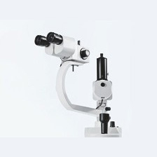
The apochromatic optics of the main microscope provide superb optical quality. The microscope image displays optimum contrast and excellent detail recognition along with a large depth of field. The bright microscope image is a particular benefit in vitreoretinal surgery. A 1:6 ratio zoom system allows the magnification of the overall system to be set as required by the surgical procedure. Two apochromatic objective lenses with focal lengths of 175 mm and 200 mm are available for different working distances. A 180° tiltable tube is used as a viewing device for the surgeon. The large tilt range allows work with minimum fatigue. The standard equipment includes eyepieces with a magnification factor of 12.5x (option: 10x). The illumination system has been designed for use in ophthalmology. An additional illumination system produces an intensive red reflex even when the eye is decentered. The angle of illumination can be switched from +2° to -2°.

Related Products
-
Hot
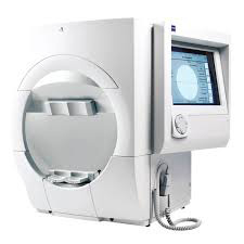
Zeiss Humphrey HFA II 750i (Pre-Owned)
Advanced analysis with comprehensive connectivity options
Validated by more than 30 years of research, design and clinical experience, the Humphrey® Field Analyzer (HFA™) is the accepted standard of care in glaucoma diagnosis and management. With over 65,000 installed units worldwide, the HFA is the premier automated visual field analyzer.- World standard of care
- All major clinical trials have relied on the Humphrey
- Connectivity to office network and EMR
- GPA™: perimetry progression software; allows for full analysis on one page
- GPA minimizes false positives by taking into account test-retest variability of the general population
- VFI™ minimizes the effects of cataracts for the purpose of measuring progression
- STATPAC™ analysis: sophisticated analysis made simple
- SITA algorithm for fast and precise visual field threshold measurements
- Gaze tracking: highly precise, real-time measurements for reliability
- SITA-SWAP provides early detection (Model 745i and 750i only)
-
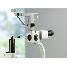
Zeiss Tonometer AT 020
The AT 020 and AT 030 Applanation Tonometers from Carl Zeiss allow precise measurement of intraocular pressure. Depending on your preference, you can attach an upright tonometer with an working position over the slit lamp swivel joint or mount a suspended tonometer on the stereomicroscope.
They were designed on the principle introduced by Professor Goldmann. This principle represents the gold standard in tonometry. Adapters allow the tonometers to be mounted to different slit lamps. Naturally, even with a mounted applanation tonometer, you still have the possibility for later slit lamp extensions.
-
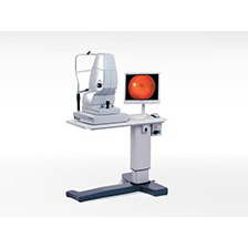
Zeiss VISUCAM NM/FA (Pre-Owned)
VISUCAM®NM/FA features legendary ZEISS optics and non-mydriatic color fundus photography enabling you to photograph through pupils as small as 3.3mm. In addition the VISUCAMNM/FA features Fluorescein Angiography (Standard) and Indocyanine Green Angiography as well as Fundus Autofluorescence capture modes (Optional). Advanced features along with an easy stereo image mode are combined with intelligent auto functions that enable reproducible and intuitive imaging for every single patient eye.
Superior patient comfort, more efficient workflow and improved eye care: The VISUCAMNM/FA from ZEISS offers many advantages. The high-quality system provides everything you need for comprehensive assessment and management of typical eye diseases such as diabetic retinopathy, glaucoma and AMD in a single workstation. -

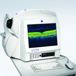
Zeiss Cirrus HD-OCT Model 400 (Pre-Owned)
The CIRRUS™ family of OCT instruments offers a choice of high-quality OCT clinical solutions to assist you in the diagnosis and management of glaucoma and retina disease across all levels of care. Focused on the essential core OCT functionality, Model 400 is designed with the smaller budget in mind. Live OCT Fundus™ technology provides the fundus image using the OCT scanner only, rather than an additional line scanning ophthalmoscope (LSO).
Cirrus HD-OCT Model 400 shares the same modern integrated design, ease of use, and small footprint as the premium performance Model 4000. The Cirrus 400 also includes; Enhanced HD rasterScan, Fast frame rate, higher transverse resolution and Anterior Segment Imaging.