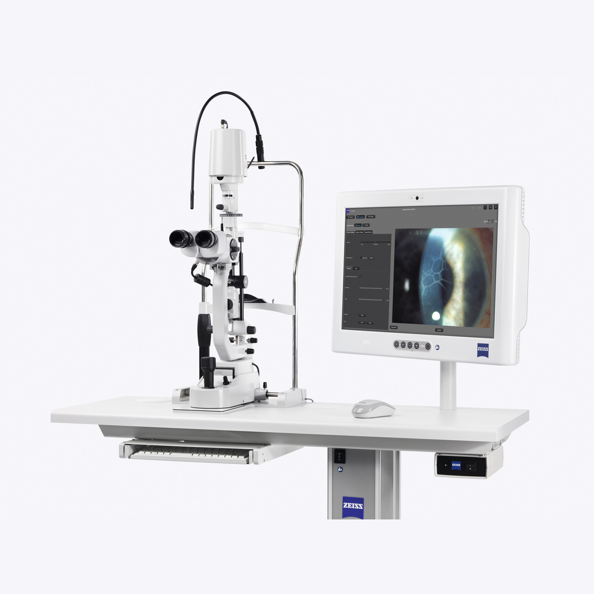
The OPMI VISU Series from Zeiss includes the VISU 150, which includes the S5 or S7 Halogen Floor stand models. This microscope has proven to be a great choice for cataract surgery. The VISU 150 provides excellent Red Reflex and multiple angles of illumination.

Related Products
-
Hot

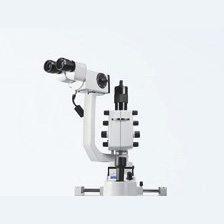
Zeiss OPMI VISU 160 Microscope (Pre-Owned)
OPMI VISU 160 As Multifaceted as the Challenges of Ophthalmic Surgery OPMI VISU® 160 was designed specifically for hospitals that perform a broad spectrum of ophthalmic surgical procedures. The versatile configuration and accessory options are tailored to meet the needs of the surgeon:
Visualization in anterior segment surgery
- With BrightFlex illumination, consisting of 2° and fading 6° illumination, for a bright red reflex and best light conditions.
- DeepView, the depth of field management system that allows you to choose between optimized depth perception or maximum light transmission.
- Integrated illuminated slit for retro-illumination during lens extraction, for example.
-
Hot
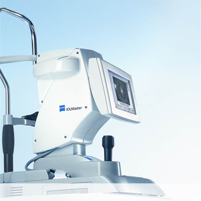
Zeiss IOL Master Model 5 (Pre-Owned)
The rapid evolution of IOL technology promises superior outcomes in cataract surgery, and it necessarily raises the bar for pre-operative biometry. The IOL Master from Carl Zeiss set the standard for highly accurate, precise measurements of all ocular characteristics necessary for IOL power calculations. The IOL Master from Carl Zeiss was the world’s first instrument for the totally non-contact measurement of all data required for the calculation of intraocular lenses. After more than ten years of clinical experience and ongoing product enhancement, the IOLMaster is truly the Gold Standard in biometry.
Two advantages for more efficiency with the IOLMaster, you save time and space in your office. All biometric measurements are possible with a single system. Without any need to relocate your patient, you measure all parameters required to calculate the IOL power: axial length, corneal radii, whiteto-white and anterior chamber depth. The results are available immediately.
-
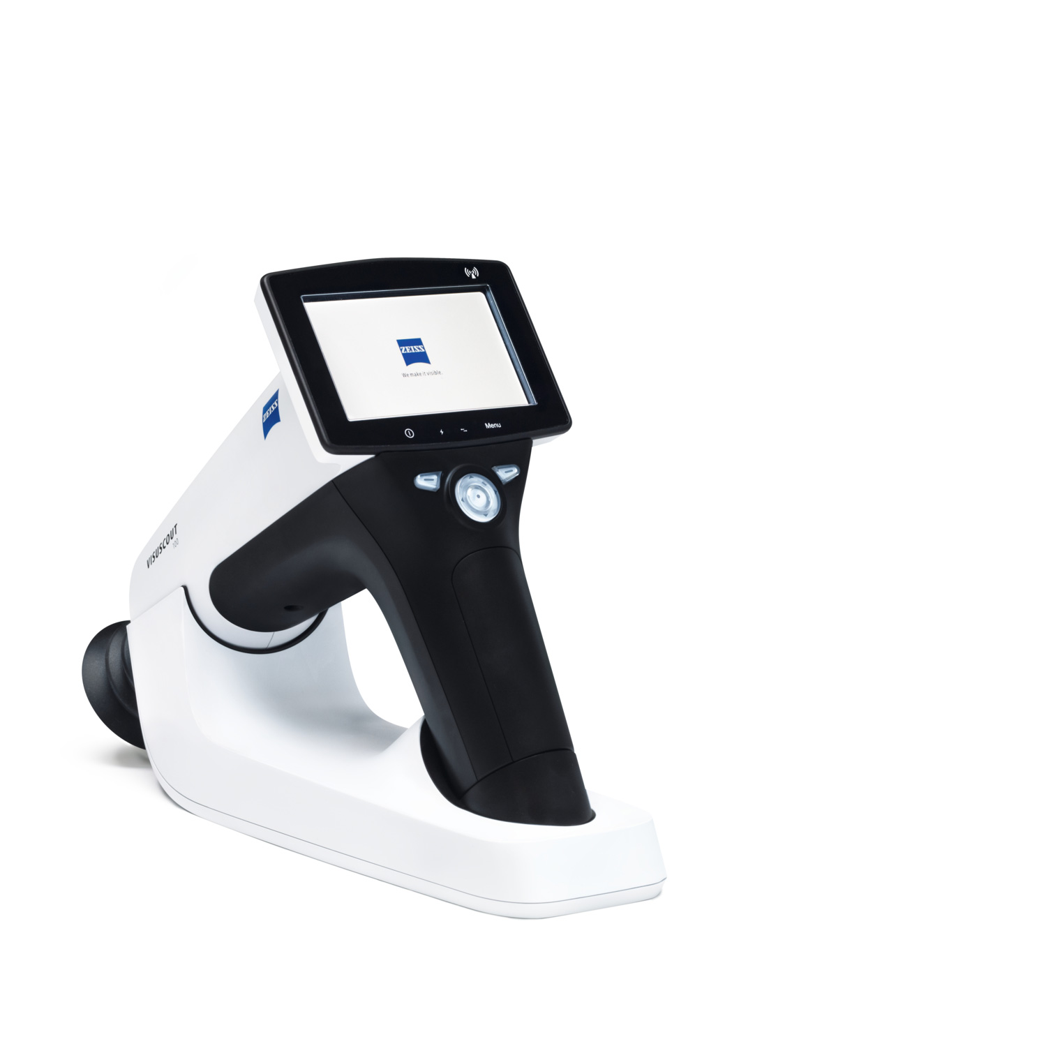
Zeiss VISUSCOUT 100 Handheld Fundus Camera
Reliably detecting and monitoring retinal disorders is key to ensuring high-quality care and to maintaining the vision of your patients. The VISUSCOUT™ 100 from ZEISS lets you do precisely that. As a mobile fundus camera, it is the perfect imaging companion.
Flexible and mobile fundus imaging
Packed into a small, rugged carrying case, the ZEISS VISUSCOUT 100 can be conveniently transported and easily fits into any practice setup. Thanks to the camera’s non-mydriatic operation and precise autofocus function, dilation of the eyes is not required. Its battery power provides added flexibility and independence. The optional WiFi functionality enables instant transfer of images to a PC or mobile device.
-
Hot
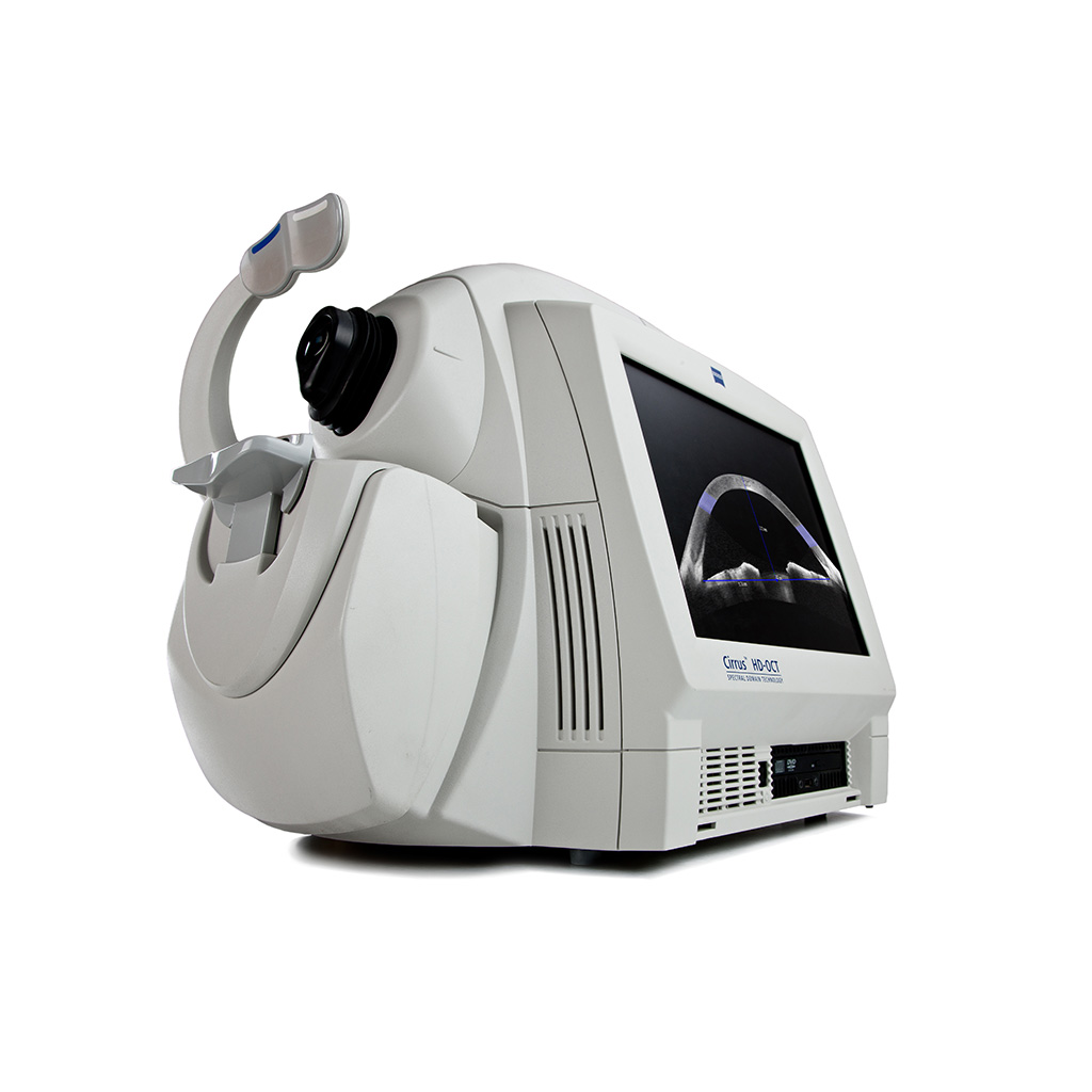
Zeiss Cirrus HD-OCT Model 4000 (Pre-Owned)
The CIRRUS™ family of OCT instruments offers a choice of high-quality OCT clinical solutions to assist you in the diagnosis and management of glaucoma and retina disease across all levels of care.
Keeping both your patients and your practice in mind, Carl Zeiss Meditec, the global leader in OCT, developed Cirrus HD-OCT. not only does it supply you with bar setting imagery, it delivers detailed diagnostic and change analysis you can rely on time and again. Along with its small footprint and fast capture speeds, Cirrus is designed to improve workflow efficiency while helping you deliver better care to your patients.