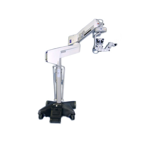
With the OPMI® VISU 210, Carl Zeiss has created a surgical microscope that can only be described as absolute state-of-the-art for ophthalmic surgery. Its wide range of uses meet the demands placed on it by surgeons and it offers new functions to improve depth of field, resolution and contrast. With its completely integrated assistant’s microscope the OPMI® VISU 210 is also ideal for university hospitals

Related Products
-

Zeiss Humphrey HFA II 740i (Pre-Owned)
Advanced analysis with comprehensive connectivity options
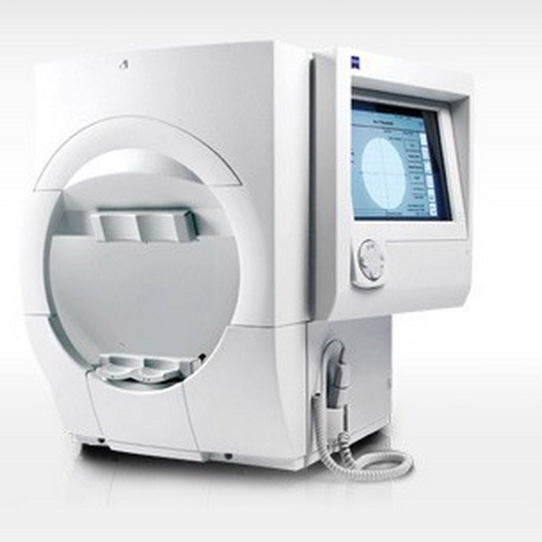
Validated by more than 30 years of research, design and clinical experience, the Humphrey® Field Analyzer (HFA™) is the accepted standard of care in glaucoma diagnosis and management. With over 65,000 installed units worldwide, the HFA is the premier automated visual field analyzer.- World standard of care
- All major clinical trials have relied on the Humphrey
- Connectivity to office network and EMR
- GPA™: perimetry progression software; allows for full analysis on one page
- GPA minimizes false positives by taking into account test-retest variability of the general population
- VFI™ minimizes the effects of cataracts for the purpose of measuring progression
- STATPAC™ analysis: sophisticated analysis made simple
- SITA algorithm for fast and precise visual field threshold measurements
- Gaze tracking: highly precise, real-time measurements for reliability
- SITA-SWAP provides early detection (Model 745i and 750i only)
-
Zeiss Humphrey HFA II 745i (Pre-Owned)
Advanced analysis with comprehensive connectivity options
Validated by more than 30 years of research, design and clinical experience, the Humphrey® Field Analyzer (HFA™) is the accepted standard of care in glaucoma diagnosis and management. With over 65,000 installed units worldwide, the HFA is the premier automated visual field analyzer.- World standard of care
- All major clinical trials have relied on the Humphrey
- Connectivity to office network and EMR
- GPA™: perimetry progression software; allows for full analysis on one page
- GPA minimizes false positives by taking into account test-retest variability of the general population
- VFI™ minimizes the effects of cataracts for the purpose of measuring progression
- STATPAC™ analysis: sophisticated analysis made simple
- SITA algorithm for fast and precise visual field threshold measurements
- Gaze tracking: highly precise, real-time measurements for reliability
- SITA-SWAP provides early detection (Model 745i and 750i only)
-
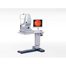
Zeiss VISUCAM NM/FA (Pre-Owned)
VISUCAM®NM/FA features legendary ZEISS optics and non-mydriatic color fundus photography enabling you to photograph through pupils as small as 3.3mm. In addition the VISUCAMNM/FA features Fluorescein Angiography (Standard) and Indocyanine Green Angiography as well as Fundus Autofluorescence capture modes (Optional). Advanced features along with an easy stereo image mode are combined with intelligent auto functions that enable reproducible and intuitive imaging for every single patient eye.
Superior patient comfort, more efficient workflow and improved eye care: The VISUCAMNM/FA from ZEISS offers many advantages. The high-quality system provides everything you need for comprehensive assessment and management of typical eye diseases such as diabetic retinopathy, glaucoma and AMD in a single workstation. -
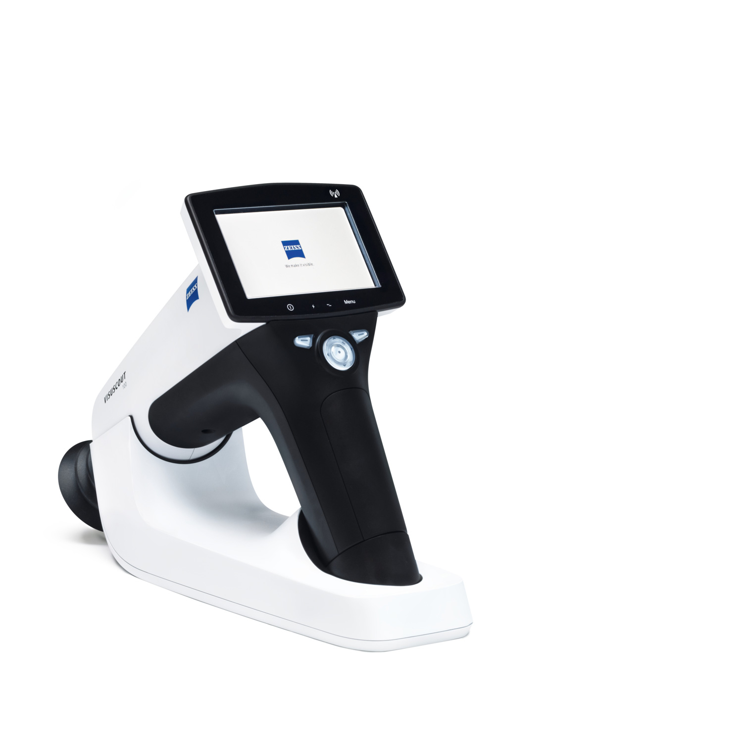
Zeiss VISUSCOUT 100 Handheld Fundus Camera
Reliably detecting and monitoring retinal disorders is key to ensuring high-quality care and to maintaining the vision of your patients. The VISUSCOUT™ 100 from ZEISS lets you do precisely that. As a mobile fundus camera, it is the perfect imaging companion.
Flexible and mobile fundus imaging
Packed into a small, rugged carrying case, the ZEISS VISUSCOUT 100 can be conveniently transported and easily fits into any practice setup. Thanks to the camera’s non-mydriatic operation and precise autofocus function, dilation of the eyes is not required. Its battery power provides added flexibility and independence. The optional WiFi functionality enables instant transfer of images to a PC or mobile device.
-
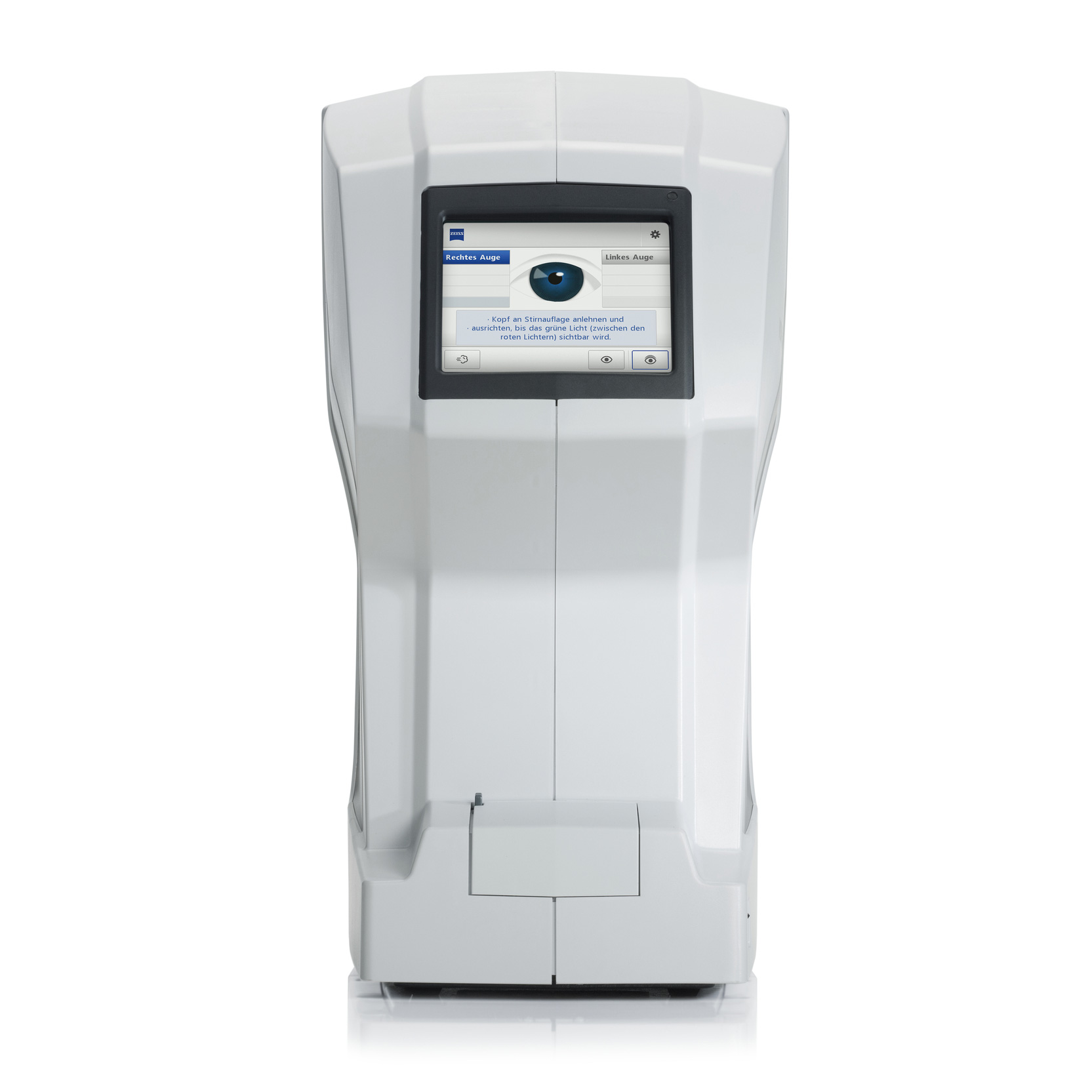
Zeiss VISUPLAN 500 Non-Contact Tonometer
The measurement of intraocular pressure is part of every professional glaucoma screening. The VISUPLAN® 500 from ZEISS makes this examination very easy and, complementing the Goldmann tonometry, does not require contact or anesthesia. The measurement is made with a soft puff of air and can be administered by your practice team.
Intuitive Operation
The measurement process runs automatically. You start directly via the touchscreen and choose from single or multiple measurements. You also have the option of initiating a test puff to prepare your patients for the examination. The results appear directly on the monitor. Alternatively, you can print out the results with the integrated thermal printer, or export via the serial interface.
-
Hot
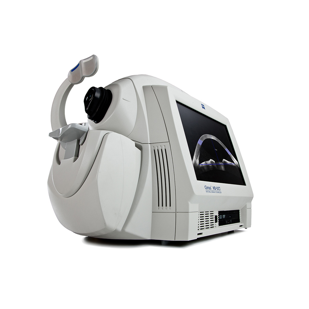
Zeiss Cirrus HD-OCT Model 4000 (Pre-Owned)
The CIRRUS™ family of OCT instruments offers a choice of high-quality OCT clinical solutions to assist you in the diagnosis and management of glaucoma and retina disease across all levels of care.
Keeping both your patients and your practice in mind, Carl Zeiss Meditec, the global leader in OCT, developed Cirrus HD-OCT. not only does it supply you with bar setting imagery, it delivers detailed diagnostic and change analysis you can rely on time and again. Along with its small footprint and fast capture speeds, Cirrus is designed to improve workflow efficiency while helping you deliver better care to your patients.