VISUCAM®NM/FA features legendary ZEISS optics and non-mydriatic color fundus photography enabling you to photograph through pupils as small as 3.3mm. In addition the VISUCAMNM/FA features Fluorescein Angiography (Standard) and Indocyanine Green Angiography as well as Fundus Autofluorescence capture modes (Optional). Advanced features along with an easy stereo image mode are combined with intelligent auto functions that enable reproducible and intuitive imaging for every single patient eye.
Superior patient comfort, more efficient workflow and improved eye care: The VISUCAMNM/FA from ZEISS offers many advantages. The high-quality system provides everything you need for comprehensive assessment and management of typical eye diseases such as diabetic retinopathy, glaucoma and AMD in a single workstation.

Zeiss VISUCAM NM/FA (Pre-Owned)
VISUCAM®NM/FA features legendary ZEISS optics and non-mydriatic color fundus photography enabling you to photograph through pupils as small as 3.3mm. In addition the VISUCAMNM/FA features Fluorescein Angiography (Standard) and Indocyanine Green Angiography as well as Fundus Autofluorescence capture modes (Optional). Advanced features along with an easy stereo image mode are combined with intelligent auto functions that enable reproducible and intuitive imaging for every single patient eye.
Superior patient comfort, more efficient workflow and improved eye care: The VISUCAMNM/FA from ZEISS offers many advantages. The high-quality system provides everything you need for comprehensive assessment and management of typical eye diseases such as diabetic retinopathy, glaucoma and AMD in a single workstation.
Zeiss VISUCAM NM/FA Specifications:
| Field angle | 45° and 30° |
| Capture modes | Color, red-frfundus ee, blue and red pictures, and pictures of the anterior segment, as well as fluorescein angiography (FA). Optional: autofluorescence, Indocyanin Green Angiography (ICG), stereo, MPOD |
| Filters | Optical filters for capture modes FA + ICG exciter and barrier filters, filters for green and blue images, filters for fundus autofluorescence and MPOD images, UV / IR barrier filters |
| Compensation for ametropia | +35 D… –35 D, continuous |
| Pupil diameter | ≥ 4.0 mm ≥ 3.3 mm (30° small pupil mode) |
| Fixation targets | External and internal (cross with 3 different sizes and hexagon) Attention mode for internal fixation target (magnified and blinking cross) Available range: +/- 15D Various programmed sequences or freely positionable as combination with stereo mode too |
| Fundus camera system | |
| Capture sequence | From 1.5 seconds (depends on flash energy) Flash steps 1-16 |
| Working distance | 40 mm ((patient eye – front lens) |
| Capture sensor | CCD 5.0 mega pixels |
| Monitor | 19“ TFT (1280 x 1024) connected via isolating transformer |
| Observation light source | 4 IR LED (each max 100mW) |
| Flash energy | Xenon flash lamp (max. 80 Ws) |
| Database | Patient information and images with field angle, FA time, R/L recognition and date of visit are stored |
| Instrument base (movement) | |
| Forward – backward | 50 mm |
| Left – right | 100 mm |
| Height | 30 mm |
| Computer / Accessories | |
| Operation system | Windows Professional |
| Hard drive | Storage of over 150,000 images possible (present size: 320 GB) |
| RAM | 1 GB |
| Interfaces | USB ports and network connectors, DVI port |
| Data interfaces | DICOM MWL, SPIF for EMR or keyboard and mouse |
| Export/import | Supported image formats: DICOM-OP, BMP, TIFF, JPEG, Patient list, DICOM MWL, DICOM storage |
| Internal DVD burner | UDF format (DVD, CD) |
| Instrument table | Asymmetric, suitable for wheelchair |
| Accessories | Network printer, USB memory stick, monitor bracket, sliding keyboard shelf for instrument table FORUM/VISUPAC archiving and image analysis system Network isolator |
| Dimensions | |
| Basic device | 410 mm x 480 mm x 650 mm (headrest) (W 16.14 x D 18.90 x H 25.59 inches) |
| Monitor | 405 x 65 x 335 mm (depends on model) (W 15.95 x D 2.56 x H 13.19 inches) |
| Weight Basic device | 30 kg (66.1 lbs) |
| Rated voltage | 100 … 240 V +/-10% (self-adjusting) |
| Frequency | 50/60 Hz |
| Power consumption | 400 VA |
Related Products
-

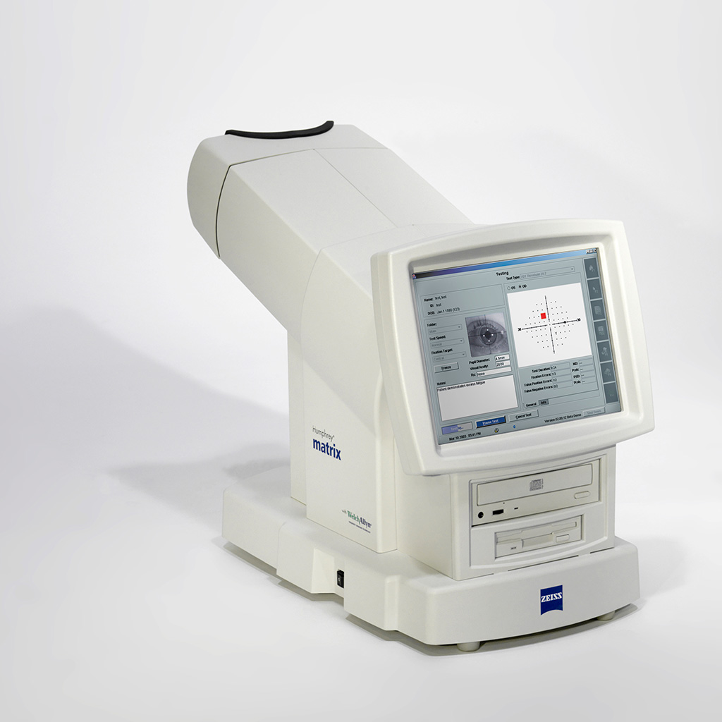
Zeiss Humphrey HFA II 740i (Pre-Owned)
Advanced analysis with comprehensive connectivity options
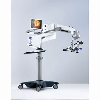
Validated by more than 30 years of research, design and clinical experience, the Humphrey® Field Analyzer (HFA™) is the accepted standard of care in glaucoma diagnosis and management. With over 65,000 installed units worldwide, the HFA is the premier automated visual field analyzer.- World standard of care
- All major clinical trials have relied on the Humphrey
- Connectivity to office network and EMR
- GPA™: perimetry progression software; allows for full analysis on one page
- GPA minimizes false positives by taking into account test-retest variability of the general population
- VFI™ minimizes the effects of cataracts for the purpose of measuring progression
- STATPAC™ analysis: sophisticated analysis made simple
- SITA algorithm for fast and precise visual field threshold measurements
- Gaze tracking: highly precise, real-time measurements for reliability
- SITA-SWAP provides early detection (Model 745i and 750i only)
-
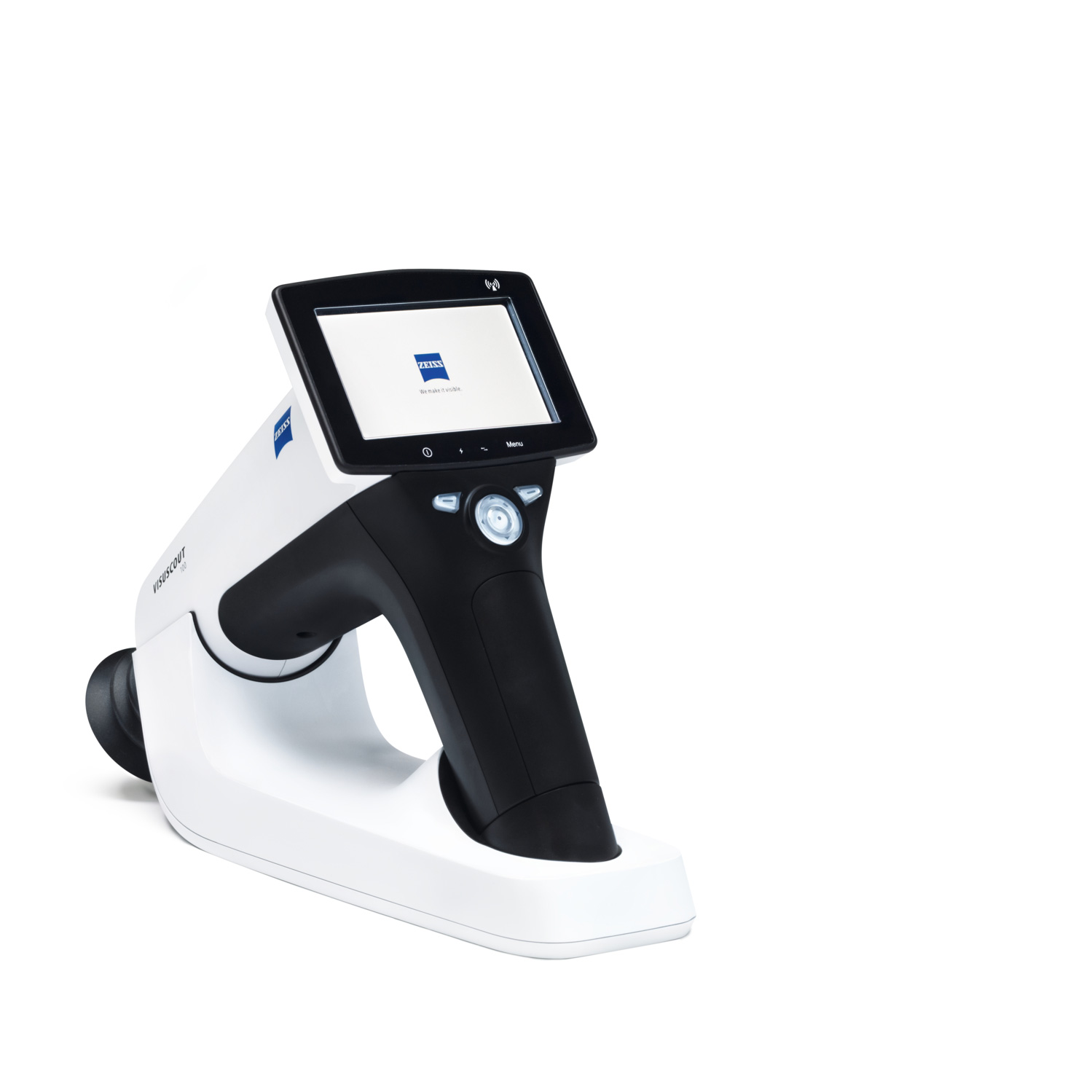
Zeiss VISUSCOUT 100 Handheld Fundus Camera
Reliably detecting and monitoring retinal disorders is key to ensuring high-quality care and to maintaining the vision of your patients. The VISUSCOUT™ 100 from ZEISS lets you do precisely that. As a mobile fundus camera, it is the perfect imaging companion.
Flexible and mobile fundus imaging
Packed into a small, rugged carrying case, the ZEISS VISUSCOUT 100 can be conveniently transported and easily fits into any practice setup. Thanks to the camera’s non-mydriatic operation and precise autofocus function, dilation of the eyes is not required. Its battery power provides added flexibility and independence. The optional WiFi functionality enables instant transfer of images to a PC or mobile device.
-
Hot
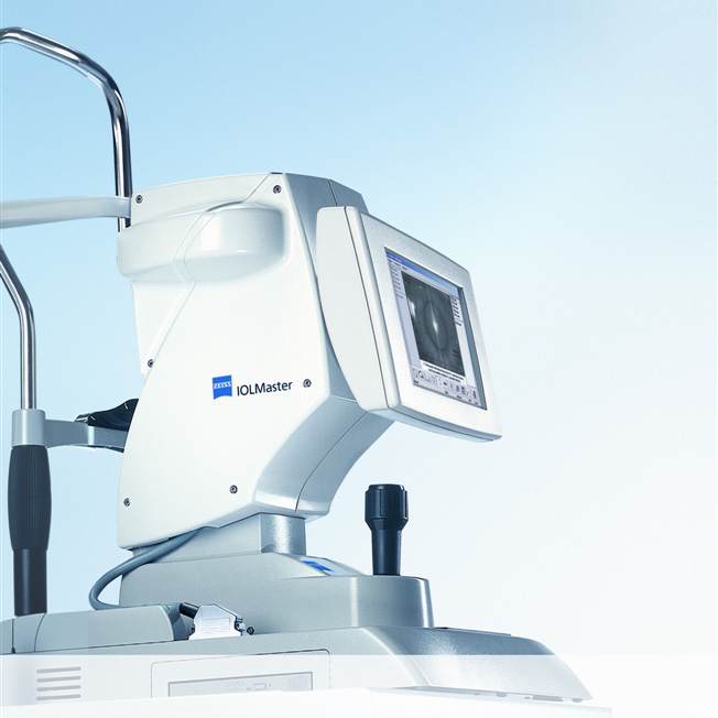
Zeiss IOL Master Model 5 (Pre-Owned)
The rapid evolution of IOL technology promises superior outcomes in cataract surgery, and it necessarily raises the bar for pre-operative biometry. The IOL Master from Carl Zeiss set the standard for highly accurate, precise measurements of all ocular characteristics necessary for IOL power calculations. The IOL Master from Carl Zeiss was the world’s first instrument for the totally non-contact measurement of all data required for the calculation of intraocular lenses. After more than ten years of clinical experience and ongoing product enhancement, the IOLMaster is truly the Gold Standard in biometry.
Two advantages for more efficiency with the IOLMaster, you save time and space in your office. All biometric measurements are possible with a single system. Without any need to relocate your patient, you measure all parameters required to calculate the IOL power: axial length, corneal radii, whiteto-white and anterior chamber depth. The results are available immediately.
-
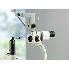
Zeiss Tonometer AT 020
The AT 020 and AT 030 Applanation Tonometers from Carl Zeiss allow precise measurement of intraocular pressure. Depending on your preference, you can attach an upright tonometer with an working position over the slit lamp swivel joint or mount a suspended tonometer on the stereomicroscope.
They were designed on the principle introduced by Professor Goldmann. This principle represents the gold standard in tonometry. Adapters allow the tonometers to be mounted to different slit lamps. Naturally, even with a mounted applanation tonometer, you still have the possibility for later slit lamp extensions.